45 draw and label the microscope
UD Virtual Compound Microscope - University of Delaware ©University of Delaware. This work is licensed under a Creative Commons Attribution-NonCommercial-NoDerivs 2.5 License.Creative Commons Attribution-NonCommercial-NoDerivs 2 Virtual Microscope - NCBioNetwork.org Lesson Description BioNetwork’s Virtual Microscope is the first fully interactive 3D scope - it’s a great practice tool to prepare you for working in a science lab. Explore topics on usage, care, terminology and then interact with a fully functional, virtual microscope. When you are ready, challenge your knowledge in the testing section to see what you have learned.
General Lab Supplies | MarketLab The Best Laboratory Supplies & Equipment MarketLab carries the lab supplies, laboratory equipment, and accessories you need to ensure your lab is running smoothly and safely. You'll find durable bins for organizing your freezers and refrigerators in the Cold Storage section. Looking for pipettes, pipette tips, and bottle top dispensers?

Draw and label the microscope
Year 7 KS3 Science Year Booklet - WordPress.com 1 Give a step by Draw a picture of a microscope. Label the eyepiece, objective lens, stage, light and focus. step instruction to a year 7 student on how to use the microscope to see a slide 2 Research a muscle cell. Draw a diagram of the cell, describe its function, describe where it is found and explain how it is adapted to its function. UD Virtual Compound Microscope - University of Delaware ©University of Delaware. This work is licensed under a Creative Commons Attribution-NonCommercial-NoDerivs 2.5 License.Creative Commons Attribution-NonCommercial-NoDerivs 2.5 License. Microscopy Pre-lab Activities - University of Delaware Microscope controls: turn knobs (click and hold on upper or lower portion of knob) throw switches (click and drag) turn dials (click and drag) move levers (click and drag) changes lenses (click and drag on objective housing) select a specimen (click on a slide)
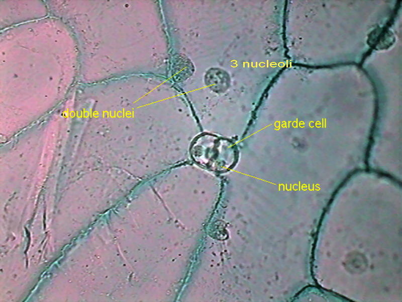
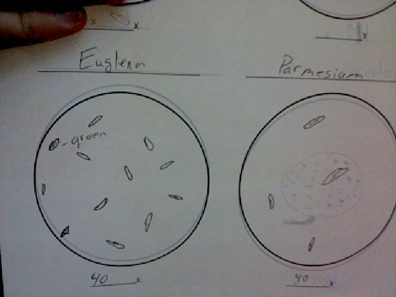
Draw and label the microscope. Pond Water Under the Microscope Here, students can sketch down what they observe and later label the different parts of the organisms. Conclusion. The primary goal here is for students to observe for themselves the different types of small organisms, which live in the pond and their diversity. Making rough sketches allows them to draw what they see and how they see them. Interactive Bacteria Cell Model - CELLS alive Periplasmic Space: This cellular compartment is found only in those bacteria that have both an outer membrane and plasma membrane (e.g. Gram negative bacteria).In the space are enzymes and other proteins that help digest and move nutrients into the cell. Cell Wall: Composed of peptidoglycan (polysaccharides + protein), the cell wall maintains the overall shape of a … Core practical 2: Use a light microscope to observe and measure ... microscope: warn against being vigorous with slides as they can splinter and advise on safe use of mounting needles. Practical techniques 4, 5 ... Draw and label the detail of these cells as accurately as you can (see fig B in the Student sheet). 10. Measure the length and breadth of two cells. Record these measurements in your diagram. Parts of a microscope with functions and labeled diagram 19/04/2022 · Q. Differentiate between a condenser and an Abbe condenser. Ans. Condensers are lenses that are used to collect and focus light from the illuminator into the specimen. They are found under the stage next to the diaphragm of the microscope. They play a major role in ensuring clear sharp images are produced with a high magnification of 400X and above.
Printable Breaker Box Electrical Panel Label Template Excel ... Password Based Circuit Breaker ... Templates For Writing Program Success Stories ... 2003 Ford Escape Fuse Box Diagram ... Electrical Transformer Symbols ... Instalasi Panel Kontrol Motor Listrik 3 Fasa ... Soil Consolidation Graph Excel ... Sheep In A Jeep Printables ... Draw And Label The Compound Microscope. PRACTICAL BOOKLET - BIOLOGY4ISC To study parts of dissecting microscope and compound microscope Physiology. Food tests : Test for starch, glucose , sucrose, protein and fats ... Draw borderlines on all four sides of 1 cm on the blank page. ... Arrows of the label should point to the part of the diagram and should not overlap each other. Microscopy Pre-lab Activities - University of Delaware Microscope controls: turn knobs (click and hold on upper or lower portion of knob) throw switches (click and drag) turn dials (click and drag) move levers (click and drag) changes lenses (click and drag on objective housing) select a specimen (click on a slide) UD Virtual Compound Microscope - University of Delaware ©University of Delaware. This work is licensed under a Creative Commons Attribution-NonCommercial-NoDerivs 2.5 License.Creative Commons Attribution-NonCommercial-NoDerivs 2.5 License.
Year 7 KS3 Science Year Booklet - WordPress.com 1 Give a step by Draw a picture of a microscope. Label the eyepiece, objective lens, stage, light and focus. step instruction to a year 7 student on how to use the microscope to see a slide 2 Research a muscle cell. Draw a diagram of the cell, describe its function, describe where it is found and explain how it is adapted to its function.
How many onion skins are there?
Fungi - Microbiology survival 101
Julia M's Bio 20 Blog: Protista

Solved: Label The Image Of Simple Columnar Epithelium Usin... | Chegg.com

The Cells and Microorganisms Webquest
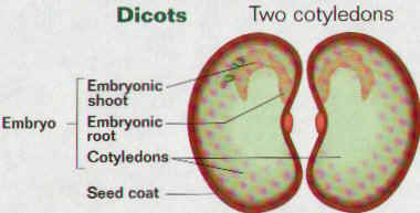
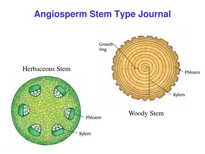
Angiosperms Biology 2 Notes
Courtney C's Bio Blog: Protista
PPT - Plant Cell Journal - Elodea PowerPoint Presentation - ID:1159196
a. Cell : unit of function - BIOLOGY4ISC
Post a Comment for "45 draw and label the microscope"